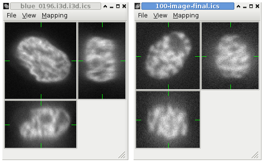
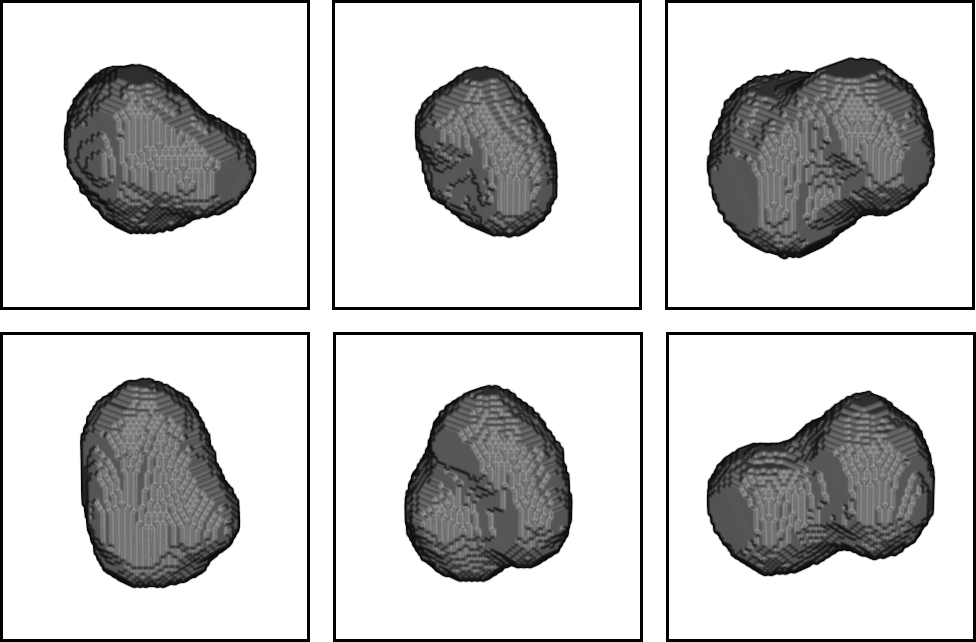
spacer.png, 0 kB
  spacer.png, 0 kB
spacer.png, 0 kB
spacer.png, 0 kB
CytoPacq: a simulation toolbox PDF Print
Written by David Svoboda   
Wednesday, 06 February 2008

Introduction

CytoPacq consists of three individual toolkits: 3D-cytogen, 3D-optigen, and 3D-acquigen. Each one is a virtual machine capable of simulating selected real processes:

  • 3D-cytogen ... a module generating the digital cell phantoms (spatial objects imitating cells and their components as well as their structure).
  • 3D-optigen ... a module simulating the transmission of the signal through the optical system.
  • 3D-acquigen ... a module simulating the phenomenons manifesting themselves during image capture with digital CCD cameras (various types of noise, sampling, digitization, etc.)

Composition of all these toolkits together into a single sequence forms a complex toolbox, which we call "CytoPacq". It covers the most common phenomenons appearing during each real acquisition process.

3D-cytogen
This module is responsible for digital phantom generation. Recently, three type of objects have been implemented: HL-60 cell nuclei, granulocyte nuclei and microspheres. However, there is no restriction put on the number of objects and the object complexity. As soon as a suitable model is defined, the corresponding digital phantom can be simply generated. The current version of 3d-cytogen supports plugins, i.e. each user can create his/her own digital phantom.

3D-optigen
The second module (3d-optigen) simulates the optical system. Namely, it involves the blurring process occurring in the optical system. Aside from blur, the devitation of the illumination axis is also taken into account.

3D-acquigen
The last stage (3d-acquigen) imitates the job of the digital CCD camera. Here, the phenomenons like noise, sampling, and quantization are simulated.


Examples

  • See some screenshots which illustrate the advance of simulation process:
    Step 1
    -> Step 2 -> Step 3 -> Step 4 -> Step 5

  • An example of four real images of (a-b) two granulocyte nuclei and (c-d) two HL60 nuclei. Each 3D figure consists of three individual images: the top-left image contains selected xy-slice, the top-right image corresponds to selected yz-slice, and the bottom one depicts selected xz-slice. Three mutually orthogonal slice planes are shown with ticks.

    (a)
     
    (b)
     
    (c)
     
    (d)

  • An example of four synthetic images of (a-b) granulocyte nuclei and (c-d) HL60 nuclei. Each 3D figure consists of three individual images: the top-left image contains selected xy-slice, the top-right image corresponds to selected yz-slice, and the bottom one depicts selected xz-slice. Three mutually orthogonal slice planes are shown with ticks.
     
    (a)
     
    (b)
     
    (c)

    (d)


Web toolkit

Currently, the version 1.0 is available. All the previous versions are listed below.
If you meet some failure or if you like to contact developers please write an e-mail to David Svoboda.

Download & Licensing

The source codes are freely available and are subject to GNU GPL. If you want to get them all you have to do is to register yourself. After that, you will obtain a free access to the download section  . Before you start compiling, building or running this application, please read the documentation carefully.

Simulated phenomenons

  • various digital phantoms available (HL60 nuclei, granulocyte nuclei, microspheres)
  • uneven illumination
  • camera selection
  • microscope selection
  • objective selection
  • dark current signal
  • (re)sampling
  • fixed pattern noise
  • quantification uncertainty (poisson noise)
  • amplifier (readout noise)
  • A/D conversion (quantization)

Papers

  • Generation of Digital Phantoms of Cell Nuclei and Simulation of Image Formation in 3D Image Cytometry, Cytometry Part A, Volume 75A, Issue 6, Pages 494-509, June 2009, ISSN:1552-4922  download
  • On Simulating 3D Fluorescent Microscope Images. In Computer Analysis of Images and Patterns. Berlin, Heidelberg: Springer-Verlag, 2007. pp 309-316, ISBN 978-3-540-74271-5 download

Presentations

  • Cytopacq: A web-based toolbox for generation of digital phantoms and simulation of image formation in 3D cell imaging, Focus On Microscopy, Krakow, Poland, April 2009 download

Credits

This service is developed and maintained by:
- David Svoboda (software development, web admin)
- Martin Maška (software development)
- Michal Kozubek (optics, signal sensing)
- Stanislav Stejskal (biology)
- Jaromír Coufal (web design)

Acknowledgement

The progress of work devoted to individual simulators was incremental in order to meet actual needs of running scientific projects No. 2B06052 and No. LC535 by the Ministry of Education of the Czech Republic.

History

  • 2009 (May)
    Main features:
    + Improved input validation.
    + Larger selection of levels of sub-pixel precision.
    + Intermediate results (images) can be downloaded while the consequentical applications are still in progress.

  • 2009 (March) - version 1.0
    Main features:
    + Enhanced list of available configurations (PSFs).
    + Larger selection of available PSFs (no need for uploading own PSF)
    + Source codes available for free after a registration.
    + All the generated images (digital phatom, ground truth, final synthetic) can be downloaded.
    + Names for all the available components amended.
    + Improved preview of the generated 3D images.

  • 2009 (January)
    Main features:
    + Error logging - in case of failure, the user is informed what had happened.
    + Flashing effect appearing during page reloading eliminated
    + Configuration used for the simulation process can be downloaded.
    + Stable solution regarding server-to-server and client-to-server communication.
    + Extended information panels for cameras, objectives and microscopes.

  • 2008 (October) - version 0.2
    Main features:
    + An improved version of web-based interface released.
    + The user can select from a larger list of cameras, objectives, microscopes, excitation and emission filters.
    + The user can watch intermediate results.

  • 2008 (May)
    Main features:
    + The first version of web-based platform-independent interface.
    + Since all the simulation steps are performed on dedicated server the user is not required to have a powerful PC.
    + A simple web-based interface as compared to the previous console-based version.
    + New digital phantoms available: granulocyte nuclei, microspheres.

  • 2007 (December) - version 0.1
    Main features:
    + This version supported only the generation of HL-60 cell nuclei.
    + Console application
    + Three individual binaries (each for one part of the simulation process) freely available  (licence enclosed) both for Win32 and Linux (kernel 2.6.x) platform.

Last Updated ( Tuesday, 09 June 2009 )
 
spacer.png, 0 kB
spacer.png, 0 kB
spacer.png, 0 kB